What Is Microscopic Endodontic Treatment? Guide & Cost
- Aileen Loo

- May 18
- 7 min read
Microscopic endodontic treatment is root canal therapy carried out under a dental operating microscope rather than with the naked eye or basic loupes. The microscope delivers up to 25 times magnification and brilliant coaxial illumination, so the dentist can see every detail of the inside of a tooth as it happens.
The result is more accurate diagnosis, cleaner canals, fewer missed problems, and a far better chance the natural tooth survives for decades. At Sola Dental in Tsim Sha Tsui, every root canal treatment is performed under a microscope as standard, not as an upgrade.

What Is Microscopic Endodontic Treatment
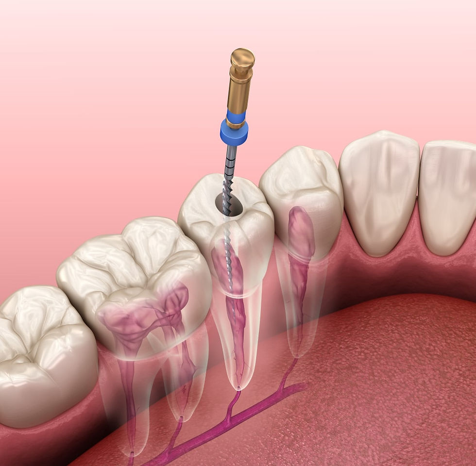
Endodontic treatment refers to any procedure performed inside the tooth on the pulp tissue, the soft core that contains nerves and blood vessels. When that pulp becomes inflamed or infected from deep decay, trauma, or repeated dental work, it must be removed, the canal disinfected, and the space sealed so bacteria cannot return.
Microscopic endodontic treatment uses a dental operating microscope, often shortened to DOM, throughout this process. The microscope sits on a flexible arm above the patient and provides:
Magnification from around 4 times up to 25 or 30 times, far beyond the 2.5 to 4.5 times offered by standard loupes
Coaxial fibre optic illumination that lights the inside of the canal along the exact line of sight
A clear, three dimensional view of canal anatomy, cracks, restorative materials, and surrounding tissues
Without this level of vision, the dentist is largely working by touch and educated guesswork. With it, every step becomes a visual decision based on what is actually inside the tooth.
Why a Microscope Matters for Root Canal Therapy
Teeth are small, the canals inside them are smaller still, and many of the structures that cause root canal failure are invisible to the unaided eye. Five clinical realities make magnification essential.
Finding Hidden and Extra Canals
Upper first molars often carry a fourth canal called the MB2, tucked under the main mesiobuccal canal. Lower molars can hide isthmuses, ribbon like connections between canals where bacteria thrive. Lower incisors regularly have two canals where most dentists treat for one.
A missed canal is the single most common reason a root canal fails years later, because infected tissue is left behind and slowly reignites the problem. Under the microscope these subtle anatomical features come into focus and can be cleaned out properly.
Diagnosing Cracks Before They Cause Failure
Vertical root fractures and hairline cracks rarely show up on standard radiographs until the damage is severe. Treating a fractured tooth as if it were simply infected wastes time and money because the fracture will keep leaking bacteria no matter how well the canal is sealed.
Under high magnification, combined with staining techniques such as methylene blue, the operator can trace the exact path of a crack and decide whether the tooth is genuinely saveable or whether extraction is the more honest answer. This protects patients from spending on procedures that have no realistic chance of success and is especially valuable for fractured teeth where the diagnosis is borderline.
Performing High Precision Microsurgery
Apical surgery, sometimes called apicoectomy, treats stubborn infection at the tip of a root when conventional retreatment is not possible. The microscope makes the difference between traditional surgery and modern endodontic microsurgery. Incisions are smaller, often only a few millimetres. Bone removal is minimised.
The root tip is resected at a controlled angle, and an ultrasonic tip preparation is filled with biocompatible material such as MTA. Adjacent nerves, sinuses, and blood vessels are clearly visible and avoided. Reported success rates for microscopic apical surgery exceed 90 percent, with noticeably less swelling and faster healing than conventional approaches.
Managing Complications Inside the Canal
Complex cases throw up complications. A file can separate inside a curved canal. A perforation can occur during access. Old posts and carriers may block the way during retreatment. Without magnification these situations often lead to extraction. Under the microscope, the operator can:
Locate a separated instrument and use specialised ultrasonic tips to loosen and remove it without destroying tooth structure
Seal perforations precisely with bioceramic materials so the canal remains usable
Remove broken posts, silver points, and old carrier based filling material safely
Carrying Out Thorough Retreatment
Endodontic retreatment is performed when a previously treated tooth becomes symptomatic or shows persistent infection on imaging. The goal is to remove every trace of old sealer and gutta percha, find any missed canals, and disinfect the entire system before refilling.
Under the microscope the dentist can tell apart restorative materials, old filling material, and natural dentine, ensuring nothing is left behind. If the original filling cannot be fully removed, the canal cannot be fully sterilised, and the retreatment is unlikely to succeed.
The Microscopic Endodontic Procedure Step by Step
Every case is tailored to the tooth in front of the dentist, but microscopic endodontic treatment follows a recognisable sequence.
Consultation and imaging. The visit begins with a full examination, dental imaging, and where needed a cone beam CT scan. This gives the operator a three dimensional map of the canal anatomy before any work begins.
Local anaesthesia and isolation. Anaesthesia is administered to numb the tooth and surrounding tissues completely. A rubber dam is placed to isolate the tooth, keep saliva away from the canal, and protect the airway.
Microscope assisted access. The microscope is positioned over the working area. The dentist creates a small access opening through the crown and uses magnification to locate every canal, including those that are calcified or hidden under the pulp chamber floor.
Cleaning and shaping. Fine nickel titanium files, often guided under the microscope, shape each canal to its working length. Irrigation with sodium hypochlorite, EDTA, and chlorhexidine flushes out bacteria and debris from areas the files cannot mechanically reach.
Disinfection check. The canal is dried with paper points and inspected under high magnification. Any remaining tissue tags, untouched walls, or extra canal openings are addressed before sealing.
Obturation. The cleaned canal is filled with a biocompatible material called gutta percha together with a sealer, creating a three dimensional seal that blocks bacteria from returning.
Restoration. A permanent restoration, usually a crown or onlay on posterior teeth, is placed to protect the tooth from fracture once it loses its pulp.
Most cases are completed in one or two visits, depending on the level of infection and the complexity of the anatomy.
Cost of Microscopic Endodontic Treatment in Hong Kong
At Sola Dental, every endodontic procedure is carried out under a dental operating microscope at no additional cost. The fee depends on which tooth is involved and whether the case is a first time root canal or a retreatment.
Procedure | Fee (HKD) |
Root Canal Treatment, Anteriors and Canines | 8,500 to 9,500 |
Root Canal Treatment, Premolars | 9,500 to 10,500 |
Root Canal Treatment, Molars | 11,000 to 12,000 |
Root Canal Retreatment, Anteriors and Canines | 11,000 to 12,000 |
Root Canal Retreatment, Premolars | 12,500 to 13,500 |
Root Canal Retreatment, Molars | 13,000 to 14,000 |
Apexification or Pulpal Regeneration | 8,500 to 10,000 |
Vital Pulp Therapy | 4,000 to 6,000 |
Apical Surgery | 9,000 to 12,000 |
Internal Bleaching | 3,000 |
Consultation | 350 |
Dental Imaging (OPG or CBCT) | 450 to 1,000 |
A final crown or onlay is usually recommended after a posterior root canal to protect the tooth and is priced at 7,500 HKD per unit. A full breakdown of clinic fees is available on the dental pricing page.
Microscopic vs Conventional Endodontics at a Glance
The microscope does not replace clinical skill. It amplifies what a skilled operator can do and turns guesswork into informed decisions.
Feature | Conventional Endodontics | Microscopic Endodontics |
Visualisation | Naked eye or 2.5x to 4.5x loupes | Up to 25x magnification with coaxial light |
Missed canal risk | Higher, especially in molars and lower incisors | Significantly reduced |
Crack diagnosis | Often impossible without surgery | Reliable using staining under magnification |
Microsurgery precision | Larger incisions, more bone removal | Minimally invasive, faster healing |
Complication management | Often leads to extraction | Perforations and separated files can frequently be repaired |
Reported success rates | 70 to 85 percent in molars | 90 percent or higher in most categories |
If a previous root canal is causing problems, an assessment under the microscope is the most reliable way to find out what is happening inside the tooth and what the realistic options are. To learn more about the team, visit the dentists page or contact the clinic directly to arrange a consultation in Tsim Sha Tsui.
FAQs About Microscopic Endodontic Treatment
Is microscopic endodontic treatment painful
No more than any other modern root canal procedure. Local anaesthesia eliminates pain during treatment, and magnification often makes the procedure quicker and more efficient, which reduces tenderness afterward. Most patients report the experience is comparable to having a large filling placed.
How long does microscopic root canal treatment take
Most single visit cases take 60 to 120 minutes. Complex molars, calcified canals, or retreatments may require two appointments spaced a week or two apart. The time spent under the microscope is what makes the difference in long term outcomes.
Why does microscopic endodontics have a higher success rate
Because the most common causes of failure, missed canals, incomplete cleaning, and undetected cracks, are exactly the problems a microscope reveals. Studies consistently report success rates of 90 percent or higher when treatment is performed under magnification, compared with 70 to 85 percent for conventional methods on molars.
Can a previously treated tooth be redone under a microscope
Yes. Retreatment under magnification is one of the strongest applications of microscopic endodontics. The dentist can remove old filling material thoroughly, find canals that were missed the first time, and seal the system again with materials that match modern standards.
Do all root canals require a microscope
Strictly speaking no, but the case for using one is overwhelming. Simple anterior cases may succeed without it, while molars, retreatments, and complex anatomy clearly benefit. At Sola Dental every endodontic procedure is performed under a microscope as standard of care, regardless of the tooth being treated.